
Abstract
Background
This study aims to compare the effects of surgical (salpingectomy) and medical (methotrexate [MTX]) treatments for ectopic pregnancy on ovarian reserve and to evaluate their relationship with anti-Müllerian hormone (AMH) and follicle-stimulating hormone (FSH) levels.
Materials and Methods
This prospective study included 76 patients diagnosed with ectopic pregnancy and a control group of 38 healthy volunteers. Patients with ectopic pregnancy were categorized into the salpingectomy (n = 38) and MTX (n = 38) groups based on clinical indications. Basal and 4-month follow-up values for AMH, FSH, and endometrial thickness were recorded. A p value of <0.05 was considered statistically significant.
Results
No statistically significant differences were found among the three groups in changes in AMH and FSH levels at the 4-month follow-up. However, FSH levels increased significantly postoperatively compared with baseline in the salpingectomy group (p < 0.05). In the MTX group, no significant change in AMH levels was observed following either single- or double-dose administration. The rate of pelvic free fluid was significantly higher in the salpingectomy group (68.4%) than in the MTX group (42.1%) (p = 0.038).
Conclusion
The findings of this study indicate that neither salpingectomy nor MTX treatments lead to a statistically significant decrease in ovarian reserve (as measured by AMH levels) at the end of a 4-month follow-up period. Although MTX treatment may offer a slight advantage in preserving hormonal balance because it carries no risk of surgical trauma or thermal injury, both treatment modalities can be considered safe in terms of future fertility.
Keywords: Ectopic pregnancy, salpingectomy, methotrexate, anti-Müllerian hormone, ovarian reserve
Introduction
The incidence of ectopic pregnancy is approximately 1-2%. The most common sites of implantation include the fallopian tubes (95%), the uterine cornua, the cervix, the ovary, the abdomen, and the previous cesarean scar (-). Today, it is one of the leading causes of maternal mortality and morbidity in the first trimester ().
In the management of ectopic pregnancy, the patient’s clinical condition, the location and size of the gestational tissue, and the desire for future fertility should be considered. Therefore, accurate diagnosis and appropriate treatment of ectopic pregnancy by contemporary obstetricians and gynecologists are essential.
Treatment options for ectopic pregnancy include expectant management, medical therapy, and surgical intervention. Expectant management, where no active treatment is applied and the patient is only monitored, is typically reserved for a limited group of patients in whom the gestational sac is not visible on ultrasound and beta-human chorionic gonadotropin (β-hCG) levels show a tendency to decline spontaneously. In medical therapy, methotrexate (MTX), a folic acid antagonist, is commonly used (). While medical management has become the most frequently chosen method due to advances in treatment modalities for ectopic pregnancy, surgical intervention remains widely practiced as well (). Surgical options include salpingectomy, salpingostomy, salpingotomy, and tubal milking. These procedures can be performed via laparotomy (L/T) or laparoscopy (L/S). The choice of surgical method depends on factors such as the patient’s age, clinical condition, surgical technique, surgeon’s experience, and available hospital resources. Mechanical and electrosurgical techniques used during surgery may negatively impact ovarian reserve due to direct vascular interruption of the ovary or thermal vascular damage caused by electrosurgery.
Ovarian reserve reflects the quantity and quality of the remaining primordial follicle pool and is an important determinant of a woman’s future reproductive potential. Among the available markers, anti-Müllerian hormone (AMH), which is secreted by the granulosa cells of preantral and small antral follicles, is regarded as one of the most reliable indicators () because it remains relatively stable throughout the menstrual cycle and is largely independent of gonadotropin fluctuations. Follicle-stimulating hormone (FSH) provides complementary information about ovarian function. Because treatment of ectopic pregnancy frequently involves the adnexa directly, surgical removal of the fallopian tube may theoretically compromise ovarian blood supply and reduce ovarian reserve, whereas systemic MTX, a folic acid antagonist, may exert a cytotoxic effect on the follicular pool. However, the available literature comparing the effects of medical and surgical treatment on ovarian reserve remains limited and inconsistent, and relatively few studies have evaluated AMH and FSH together against a healthy control group (,). Clarifying whether these two widely used treatment modalities differ in their effect on ovarian reserve is therefore clinically relevant when counseling women who wish to preserve future fertility.
The aim of this study was to compare the effects of surgical (salpingectomy) and medical (MTX) treatments of ectopic pregnancy on ovarian reserve, as assessed by AMH and FSH levels, in women of reproductive age who wished to preserve future fertility.
Materials and Methods
This prospective study was conducted at the Obstetrics and Gynecology Clinic of Bursa Yüksek İhtisas Training and Research Hospital, affiliated with the University of Health Sciences Türkiye. The study was approved by Bursa Yüksek İhtisas Training and Research Hospital Ethics Committee under protocol number 2011-KAEK-25 2022/10-12 (date: 05.10.2022). This study was conducted as part of the corresponding author’s medical specialty thesis while working at Bursa Yüksek İhtisas Training and Research Hospital. Ethical approval was obtained from the same institution during the study period. It included 76 patients diagnosed with ectopic pregnancy and a control group of 38, spanning one year beginning on October 6, 2022. Written and verbal informed consent was obtained from all participants. The sample size for the control group (n = 38) was determined based on a power analysis conducted using G*Power software, aiming for 80% power and a 5% significance level to detect a moderate effect size between the groups. Additionally, the patients were informed about the study by the obstetrician conducting the research.
Among the included patients, data such as age, weight, height, body mass index (BMI), gravidity, parity, history of abortion, β-hCG levels, AMH levels (electrochemiluminescence immunoassay [ECLIA] method), FSH levels, endometrial thickness, adnexal mass size, and presence of free pelvic fluid were recorded. As medical treatment, MTX 50 mg/m2 was administered intramuscularly. Blood tests were performed on days 4 and 7. In 24 patients who received MTX treatment, a second dose of MTX was administered on day 7 because follow-up tests showed less than a 15% decrease in β-hCG levels. Patients were not randomized; instead, they were allocated to either the surgical or the medical treatment group based on clinical indications, such as hemodynamic stability, β-hCG levels, and the presence of tubal rupture, in accordance with standard clinical guidelines.
The inclusion criteria for the study were as follows: patients aged between 18 and 45 years; diagnosed with ectopic pregnancy and hospitalized for surgical treatment of the condition; without additional comorbidities, and willing to participate in the study. Patients who were outside the 18-45 age range, who had existing comorbid conditions, who had previously undergone procedures that could affect ovarian reserve, or who were unwilling to participate were excluded from the study.
The control group consisted of 38 healthy volunteers who presented to the gynecology outpatient clinic during the same study period and had no current or recent pregnancy, no history of ectopic pregnancy, and no previous surgery or medical condition known to affect ovarian reserve. Control participants were selected to match the treatment groups in reproductive age (18-45 years) and were required to have regular menstrual cycles and not to have used hormonal contraception or ovulation-inducing agents in the preceding three months. The same inclusion and exclusion criteria with respect to comorbidities and prior procedures affecting the ovary were applied to the control group.
Follow-up Protocol and Assessment of Ovarian Reserve
Ovarian reserve was assessed in all three groups at baseline (at the time of diagnosis, before any treatment) and again at the fourth month of follow-up. AMH was measured in serum using the ECLIA, and FSH was measured in the early follicular phase. Endometrial thickness was evaluated by the same examiner using transvaginal ultrasonography. Endometrial thickness was recorded because it reflects the endometrial response to the prevailing hormonal milieu and the resolution of the gestational process, and was monitored to document the return of the endometrium to a non-pregnant baseline after treatment. In the MTX group, β-hCG was monitored on the fourth and seventh days; a second MTX dose was administered on the seventh day, when the decrease in β-hCG was less than 15%. The fourth-month visit was selected as the primary follow-up time point because it allows sufficient time for the acute physiological effects of pregnancy and treatment to subside, and for AMH levels to reflect a stable, non-pregnant ovarian state.
Study Outcomes
The primary outcome of the study was the change in ovarian reserve, defined as the change in serum AMH level from baseline to the fourth month of follow-up, and was compared among the salpingectomy, MTX, and control groups. The secondary outcomes were the corresponding changes in FSH levels and in endometrial thickness over the same period.
Statistical Analysis
The variables were expressed as descriptive statistics using mean ± standard deviation, median (minimum-maximum), and frequency (percentage) values. The assumption of normality was assessed with the Shapiro-Wilk test. For comparisons of normally distributed quantitative variables between three groups, the one-way analysis of variance test was used, and for comparisons between two groups, the Student’s t-test was applied. For non-normally distributed quantitative variables, the Kruskal-Wallis test was used for comparisons among three groups, and the Mann-Whitney U test was used for comparisons between two groups. The chi-square test or Fisher’s exact test was used to compare categorical variables. The distributions of baseline and 4-month AMH and FSH values and of endometrial thickness across groups were illustrated using box plots. Additionally, changes in AMH, FSH, and endometrial thickness between baseline and the 4th month within groups and among subgroups stratified by MTX dosage were analyzed using the Wilcoxon test.
The analyses were conducted using IBM SPSS Statistics, version 22.0 (IBM Corp., Armonk, NY, USA), with a significance level of 0.05.
Results
A total of 114 patients were included in the study and were divided into three groups: the salpingectomy group (n = 38), the MTX group (n = 38), and the control group (n = 38). The demographic and obstetric characteristics of the study groups are presented in . As shown in the table, there were no statistically significant differences among the three groups with respect to age, height, weight, BMI, parity and the numbers of vaginal deliveries, cesarean sections, and abortions. However, a statistically significant difference in gravidity was observed in at least one group.
Table 1. Demographic and obstetric characteristics of the salpingectomy, methotrexate, and control groups.
Characteristics | Salpingectomy group (n = 38) | Methotrexate group (n = 38) | Control group (n = 38) | p value |
|---|---|---|---|---|
Age (years) | 32.1 ± 5.3 | 30.4 ± 5.3 | 28.9 ± 5.9 | 0.053 |
Height (cm) | 160.6 ± 5.23 | 161.8 ± 4.99 | 161.1 ± 5.84 | 0.615 |
Weight (kg) | 68.5 (45-82) | 67 (51-105) | 68 (55-88) | 0.892 |
BMI (kg/m2) | 26.1 (19.2-34.1) | 25.5 (20.96-36.3) | 26.2 (21.3-35.7) | 0.827 |
Gravidity (n) | 3 (1-9) | 3 (1-7) | 1 (0-4) | <0.001 |
Parity (n) | 1 (0-8) | 1 (0-6) | 1 (0-3) | 0.397 |
Number of vaginal births | 0 (0-8) | 0 (0-4) | 0 (0-3) | 0.483 |
Number of cesarean sections | 0 (0-5) | 0 (0-3) | 0 (0-3) | 0.960 |
Number of abortions | 0 (0-2) | 0 (0-3) | 0 (0-2) | 0.440 |
Kruskal-Wallis test. BMI, body mass index.
The laboratory and sonographic characteristics of the salpingectomy, MTX, and control groups are presented in . No statistically significant differences were observed among the three groups with respect to changes in AMH, FSH levels at the 4th month, and control endometrial thickness. However, baseline AMH, 4-month AMH, baseline FSH, changes in FSH, baseline endometrial thickness, and changes in endometrial thickness showed statistically significant differences in at least one of the groups (p < 0.05). The distributions of AMH and FSH changes are shown in and , respectively.
Table 2. Laboratory and sonographic characteristics of the salpingectomy, methotrexate, and control groups.
- | Salpingectomy group (n=38) | Methotrexate group (n=38) | Control group (n=38) | p |
|---|---|---|---|---|
Initial AMH (ng/mL) | 1.53 (0.25-5.1) | 1.84 (0.17-6.5) | 2.61 (0.27-8.25) | 0.006 |
4th month AMH (ng/mL) | 1.36 (0.3-4.4) | 1.99 (0.18-6.84) | 2.4 (0.29-8.1) | 0.012 |
AMH change | 0.01 (-0.79-1.09) | 0.07 (-0.63-1.34) | -0.12 (-0.57-0.82) | 0.568 |
Initial FSH (mIU/mL) | 1.06 (0.05-6.2) | 2.35 (0.11-10.95) | 5.54 (2.6-9.1) | <0.001 |
4th month FSH (mIU/mL) | 5.72 ± 2.1 | 5.9 ± 2.02 | 5.9 ± 2.03 | 0.935 |
FSH change | 4.07 (-1.20-9.05) | 3.36 (-6.77-9.23) | 0.59 (-5.51-6.51) | <0.001 |
Initial endometrial thickness (mm) | 10.5 (4-23) | 8 (4-25) | 5 (2-8) | <0.001 |
Control endometrial thickness (mm) | 6 (2-11) | 5 (3-11) | 6 (2-10) | 0.607 |
Endometrial thickness change | -4.5 (-15-1) | -2 (-15-4) | 0 (-6-5) | <0.001 |
Kruskal-Wallis test. AMH, anti-Müllerian hormone; FSH, follicle-stimulating hormone.
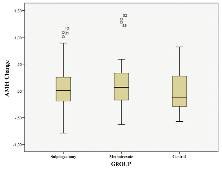
Box plot illustrating the distribution of anti-Müllerian hormone (AMH) changes in the salpingectomy, methotrexate, and control groups.
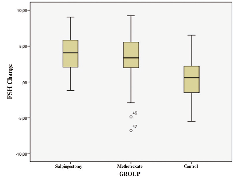
Box plot illustrating the distribution of follicle-stimulating hormone (FSH) changes in the salpingectomy, methotrexate, and control groups.
The pairwise comparisons among the salpingectomy, MTX, and control groups are presented in . No significant differences were observed between the salpingectomy and MTX groups with respect to gravida and baseline FSH values. However, there were statistically significant differences between the salpingectomy and control groups and between the MTX and control groups. When evaluating baseline and 4-month AMH values, a significant difference was found only between the salpingectomy and control groups. In contrast, baseline FSH values, FSH changes, and endometrial thickness changes showed statistically significant differences between the salpingectomy and control groups, and between the MTX and control groups. Baseline endometrial thickness exhibited statistically significant differences across all groups.
Table 3. Bivariate evaluation of the salpingectomy, methotrexate, and control groups.
Group | p | |
|---|---|---|
Gravida | Salpingectomy-methotrexate Salpingectomy-control Methotrexate-control | 1.000 <0.001 <0.001 |
Initial AMH | Salpingectomy-methotrexate Salpingectomy-control Methotrexate-control | 0.411 0.004 0.258 |
4th month AMH | Salpingectomy-methotrexate Salpingectomy-control Methotrexate-control | 0.379 0.009 0.449 |
Initial FSH | Salpingectomy-methotrexate Salpingectomy-control Methotrexate-control | 0.415 <0.001 <0.001 |
FSH change | Salpingectomy-methotrexate Salpingectomy-control Methotrexate-control | 1.000 <0.001 <0.001 |
Initial endometrial thickness | Salpingectomy-methotrexate Salpingectomy-control Methotrexate-control | 0.022 <0.001 <0.001 |
Endometrial thickness change | Salpingectomy-methotrexate Salpingectomy-control Methotrexate-control | 0.085 <0.001 0.001 |
Mann-Whitney U test. AMH, anti-Müllerian hormone; FSH, follicle-stimulating hormone.
In the salpingectomy group, the median β-hCG level was 3626.5 (range: 207–61500), while in the MTX group it was 1373.5 (range: 66–13100). An adnexal mass was present in 36 patients (94.7%) in the salpingectomy group, with a median mass size of 25.5 mm (range: 6–60 mm). In the MTX group, an adnexal was detected in 32 patients (84.2%), with a median size of 20.5 mm (range: 7–70 mm). There were no statistically significant differences between the two groups in the presence or size of the adnexal mass (p = 0.262 and p = 0.635, respectively). Free fluid was observed in 68.4% (21 patients) of the salpingectomy group and 42.1% (16 patients) of the MTX group, representing a statistically significant difference between the groups (p = 0.038).
Preoperative (baseline) and postoperative (4-month) AMH, FSH, and endometrial thickness were compared within each group; the results for all three groups are summarized in . AMH did not change significantly in any group. In contrast, FSH increased significantly after treatment in both the salpingectomy and MTX groups, and endometrial thickness decreased significantly in both treatment groups. No significant changes were observed in the control group. Additional subgroup analyses are presented in the .
Table 4. Comparison of pre-treatment/baseline and post-treatment/4th-month findings within the salpingectomy, methotrexate, and control groups.
Parameter | Pre-treatment/baseline | Post-treatment/4th month | p |
|---|---|---|---|
Salpingectomy group (n = 38) | |||
AMH (ng/mL) | 1.53 (0.25-5.1) | 1.36 (0.3-4.4) | 0.572 |
FSH (mIU/mL) | 1.06 (0.05-6.2) | 5.41 (1.70-10.1) | <0.001 |
Endometrial thickness (mm) | 10.5 (4-23) | 6 (2-11) | <0.001 |
Methotrexate group (n = 38) | |||
AMH (ng/mL) | 1.84 (0.17-6.5) | 1.99 (0.18-6.84) | 0.313 |
FSH (mIU/mL) | 2.35 (0.11-10.95) | 5.75 (2.3-9.81) | <0.001 |
Endometrial thickness (mm) | 8 (4-25) | 5 (3-11) | <0.001 |
Control group (n = 38) | |||
AMH (ng/mL) | 2.61 (0.27-8.25) | 2.4 (0.29-8.1) | 0.772 |
FSH (mIU/mL) | 5.54 (2.6-9.1) | 6 (1.7-9.81) | 0.429 |
Endometrial thickness (mm) | 5 (2-8) | 6 (2-10) | 0.272 |
Wilcoxon signed-rank test. AMH, anti-Müllerian hormone; FSH, follicle-stimulating hormone.
Discussion
The most striking finding of our study is that, although both MTX and salpingectomy are effective and safe for future fertility, MTX better preserves AMH levels than surgical intervention. While previous studies have often debated the impact of these treatments on ovarian reserve, our findings highlight a clear divergence in the biological response between systemic medical therapy and localized surgical trauma.
Our observation that MTX does not significantly impair ovarian reserve is consistent with the findings of Oriol et al. () and Shirazi et al. (), who reported that systemic administration of MTX does not lead to a long-term decline in AMH (). However, our study goes a step further by directly comparing these results with those of a surgical cohort under similar physiological conditions. Unlike the MTX group, the salpingectomy group in our study exhibited a significant postoperative rise in FSH levels. The potential effects of salpingectomy on ovarian reserve have been examined in previous studies (,). By comparing these two modalities, we demonstrate that the surgical approach—specifically the use of electrosurgical tools—presents a higher risk of localized vascular compromise than the non-invasive MTX approach.
The biological explanation for the preservation of AMH in the MTX group, even in cases requiring double doses, may lie in the unique hormonal environment of pregnancy. High levels of β-hCG and the suppressed FSH levels characteristic of the first trimester likely provide a “protective shield” for the primordial follicle pool against the cytotoxic effects of folic acid antagonists (-). In contrast, the rise in FSH observed in our surgical group likely stems from the interruption of the ovarian blood supply. Even with collateral circulation from the internal iliac artery, the mesosalpingeal damage during salpingectomy can impair ovarian microcirculation, a phenomenon that has been suggested in literature (,) but is clearly evidenced by the hormonal shifts in our patient cohort.
Furthermore, our data revealed that AMH levels tended to stabilize or slightly increase during the four-month follow-up period in all groups. This observation contrasts with some earlier reports that suggested a permanent decline after salpingectomy (,-). This discrepancy can be explained by recovery of granulosa cell activity once the suppressive effect of pregnancy and the immediate inflammatory response to surgery subside. Thus, while the striking difference is visible in the immediate post-treatment period, the long-term fertility outlook remains positive for both groups.
A notable observation in our cohort was that the control group had higher baseline AMH and FSH values than the two treatment groups. Several factors likely contribute to these differences. First, the groups were not randomized; patients were allocated to surgical or medical treatment based on clinical indications, which may have introduced differences in age and reproductive history between the groups. Consistent with this finding, gravidity differed significantly between the control and treatment groups, suggesting differences in reproductive histories. Second, baseline samples from the ectopic pregnancy groups were obtained during an ongoing hormonally active gestation when elevated β-hCG and the suppressive effect of pregnancy on the hypothalamic-pituitary-ovarian axis can transiently lower measured FSH and may also modestly influence circulating AMH (,), whereas control measurements were obtained in a non-pregnant state. These physiological and selection-related factors, rather than a true difference in underlying ovarian reserve, are the most plausible explanation for the baseline discrepancies. Because our primary analysis focused on the within-group change from baseline to the fourth month rather than on absolute between-group values, the conclusions regarding the effect of treatment on ovarian reserve are not invalidated by these baseline differences.
Endometrial thickness was included as a secondary parameter because it serves as an indirect measure of the patient’s hormonal and gestational status rather than a direct measure of ovarian reserve. The significantly greater baseline endometrial thickness in the treatment groups compared with controls is consistent with the decidual response associated with an ongoing pregnancy, and the significant decrease observed at follow-up in both the salpingectomy and MTX groups reflects the expected regression of the endometrium following resolution of the pregnancy. Importantly, endometrial thickness returned to values comparable to those in the control group by the fourth month, with no significant between-group difference observed at follow-up. This finding indicates that neither treatment modality had a lasting adverse effect on the endometrium and that the uterine environment recovers similarly after both medical and surgical management. Endometrial thickness should therefore be regarded as a supportive marker of post-treatment recovery rather than a primary determinant of the study’s main conclusions, which rest on the AMH-based assessment of ovarian reserve.
Taken together, our study provides a comparative perspective that was previously lacking in the literature. By contrasting the mechanical impact of surgery with the systemic safety of MTX, we demonstrate that MTX should be the preferred option when preservation of the immediate hormonal balance of the ovarian reserve is a clinical priority.
Study Limitations
One limitation of our study is the lack of uniformity in surgical management because not all salpingectomy procedures were performed by the same surgical team, and this may have affected the consistency of outcome assessment. Further limitations include the single-center, non-randomized design in which patients were allocated to treatment based on clinical indications; this may have introduced selection bias and contributed to the baseline differences observed between groups. The relatively modest sample size and the limited four-month follow-up period also restrict the ability to detect small or delayed changes in ovarian reserve and to evaluate longer-term fertility outcomes. Despite these limitations, our study has several strengths. To our knowledge, it is one of the few prospective studies that directly compare the effects of surgical and medical treatments for ectopic pregnancy on ovarian reserve within the same time frame and include a healthy control group as a reference. The simultaneous and serial evaluation of multiple complementary markers of ovarian function, namely AMH and FSH, together with paired baseline and follow-up measurements, strengthens the internal validity of the comparison and provides a more comprehensive assessment of ovarian reserve than a single marker.
Conclusion
Our study demonstrates that neither salpingectomy nor MTX treatment for ectopic pregnancy results in a statistically significant reduction in ovarian reserve (as measured by AMH levels) after four months. Although MTX appears to preserve AMH levels slightly better, which is likely due to the absence of surgical thermal damage, both modalities remain safe with respect to future fertility. These findings should be confirmed by larger, multi-center studies with longer follow-up periods.
Ethics
Ethics Committee Approval: Bursa Yüksek İhtisas Training and Research Hospital Ethics Committee under protocol number 2011-KAEK-25 2022/10-12 (date: 05.10.2022). This study was conducted as part of the corresponding author’s medical specialty thesis while working at Bursa Yüksek İhtisas Training and Research Hospital. Ethical approval was obtained from the same institution during the study period.
Informed Consent: Written and verbal informed consent was obtained from all participants.
Footnotes
Authorship Contributions
Surgical and Medical Practices: B.T.K., D.Ş., Concept: B.A.S., Z.S., Design: B.T.K., D.Ş., Data Collection or Processing: B.T.K., B.A.S., Z.S., D.Ş., Analysis or Interpretation: B.T.K., B.A.S., Z.S., D.Ş., Literature Search: B.T.K., Writing: B.T.K.
Conflict of Interest
No conflict of interest was declared by the authors.
Financial Disclosure
The authors declared that this study received no financial support.
References
Ectopic pregnancy—United States, 1990-1992. MMWR Morb Mortal Wkly Rep.
Van Den Eeden SK, Shan J, Bruce C, Glasser M. Ectopic pregnancy rate and treatment utilization in a large managed care organization. Obstet Gynecol. 2005;105:1052-1057.
American College of Obstetricians and Gynecologists’ Committee on Practice Bulletins—Gynecology. ACOG Practice Bulletin No. 193: Tubal Ectopic Pregnancy. Obstet Gynecol. 2018;131:e91-e103. Erratum in: Obstet Gynecol. 2019;133:1059.
Ory SJ, Nnadi E, Herrmann R, O’Brien PS, Melton LJ 3rd. Fertility after ectopic pregnancy. Fertil Steril. 1993;60:231-235.
Po L, Thomas J, Mills K, Zakhari A, Tulandi T, Shuman M, et al. Guideline no. 414: management of pregnancy of unknown location and tubal and nontubal ectopic pregnancies. J Obstet Gynaecol Can. 2021;43:614-630.e1.
Tanaka T, Hayashi H, Kutsuzawa T, Fujimoto S, Ichinoe K. Treatment of interstitial ectopic pregnancy with methotrexate: report of a successful case. Fertil Steril. 1982;37:851-852.
Practice Committee of American Society for Reproductive Medicine. Medical treatment of ectopic pregnancy: a committee opinion. Fertil Steril. 2013;100:638-644.
de Vet A, Laven JS, de Jong FH, Themmen AP, Fauser BC. Antimüllerian hormone serum levels: a putative marker for ovarian aging. Fertil Steril. 2002;77:357-362.
Askari E. A comparative study of impacts of single-dose systemic methotrexate and salpingectomy surgery treatments on ovarian reserve in women with ectopic pregnancy using anti-Mullerian hormone. Asian J Pharm. 2018;12:S304-S308.
Luo J, Shi Y, Liu D, Yang D, Wu J, Cao L, et al. The effect of salpingectomy on the ovarian reserve and ovarian response in ectopic pregnancy: a systematic review and meta-analysis. Medicine (Baltimore). 2019;98:e17901.
Oriol B, Barrio A, Pacheco A, Serna J, Zuzuarregui JL, Garcia-Velasco JA. Systemic methotrexate to treat ectopic pregnancy does not affect ovarian reserve. Fertil Steril. 2008;90:1579-1582.
Shirazi M, Pooransari P, Hajiha N, Shaker Z, Ghazi M, Davari Tanha F, et al. Effect of single-dose methotrexate treatment on ovarian reserve in women with ectopic pregnancy undergoing infertility treatment: a single-center experience. Int J Fertil Steril. 2020;14:23-26.
Hill MJ, Cooper JC, Levy G, Alford C, Richter KS, DeCherney AH, et al. Ovarian reserve and subsequent assisted reproduction outcomes after methotrexate therapy for ectopic pregnancy or pregnancy of unknown location. Fertil Steril. 2014;101:413-41s9.
Rodgers R, Carter J, Reid G, Krishnan S, Ludlow J, Cooper M, et al. The effect of laparoscopic salpingectomy for ectopic pregnancy on ovarian reserve. Aust N Z J Obstet Gynaecol. 2020;60:278-283.
Grynnerup AG, Lindhard A, Sørensen S, Ørskov M, Petersen KR, Madsen LB, et al. Serum anti-Müllerian hormone concentration before and after salpingectomy for ectopic pregnancy. Reprod Biomed Online. 2018;37:624-630.
Ulug P, Oner G. Evaluation of the effects of single or multiple dose methotrexate administration, salpingectomy on ovarian reserve of rat with the measurement of anti-Müllerian hormone (AMH) levels and histological analysis. Eur J Obstet Gynecol Reprod Biol. 2014;181:205-209.
Nelson SM, Stewart F, Fleming R, Freeman DJ. Longitudinal assessment of antimüllerian hormone during pregnancy-relationship with maternal adiposity, insulin, and adiponectin. Fertil Steril. 2010;93(4):1356-1358.
Pankhurst MW, Clark CA, Zarek J, Laskin CA, McLennan IS. Changes in circulating ProAMH and total AMH during healthy pregnancy and post-partum: a longitudinal study. PLoS One. 2016;11:e0162509.
Wang S, Gu J. The effect of prophylactic bilateral salpingectomy on ovarian reserve in patients who underwent laparoscopic hysterectomy. J Ovarian Res. 2021;14:86.
Findley AD, Siedhoff MT, Hobbs KA, Steege JF, Carey ET, McCall CA, et al. Short-term effects of salpingectomy during laparoscopic hysterectomy on ovarian reserve: a pilot randomized controlled trial. Fertil Steril. 2013;100:1704-1708.
Yuan Z, Cao D, Bi X, Yu M, Yang J, Shen K. The effects of hysterectomy with bilateral salpingectomy on ovarian reserve. Int J Gynaecol Obstet. 2019;145:233-238.
Galanderov R, Kürek Eken M, İlhan G, Keskin M, Turfanda A. Evaluation of ovarian reserve using anti-Müllerian hormone and ultrasonography in patients undergoing hysterectomy. Istanbul Med J. 2015;78:67-71.
Özşener S, Şimşek D. Effect of bilateral salpingectomy on ovarian reserve in patients undergoing hysterectomy for benign conditions [master’s thesis]. İzmir: Ege University, Faculty of Medicine; 2016.
Supplementary Materials
Supplementary Materials
Download